Researchers at the All India Institute of Medical Sciences (AIIMS), New Delhi, have identified two critical anatomical regions that require heightened surgical caution during complex cancer procedures. The findings aim to support safer surgical techniques by reducing the risk of excessive bleeding and accidental damage to vital organs and blood vessels.
Dr M D Ray, Professor in the Department of Surgical Oncology at Dr BRA-IRCH, AIIMS Delhi, and his team have described two surgical reference areas, known as “Ray’s Zone” in the pelvis and “Ray’s Hepatocaval Triangle” near the liver. Both concepts focus on identifying high-risk anatomical structures before surgical dissection.
Ray’s Zone Identified During Gynaecological Cancer Surgeries
The researchers identified a region below the uterine vessels, termed the “Red Alert Zone” or “Ray’s Zone”, where several important anatomical structures are located close to one another. These include the ureter, urinary bladder, pelvic nerves and major blood vessels.
The concept was developed following an analysis of 255 radical and modified radical hysterectomy procedures performed on patients with cervical, endometrial and ovarian cancers.
After examining surgical anatomy and factors associated with bleeding and organ injuries, the team modified its surgical approach in the later 105 cases.
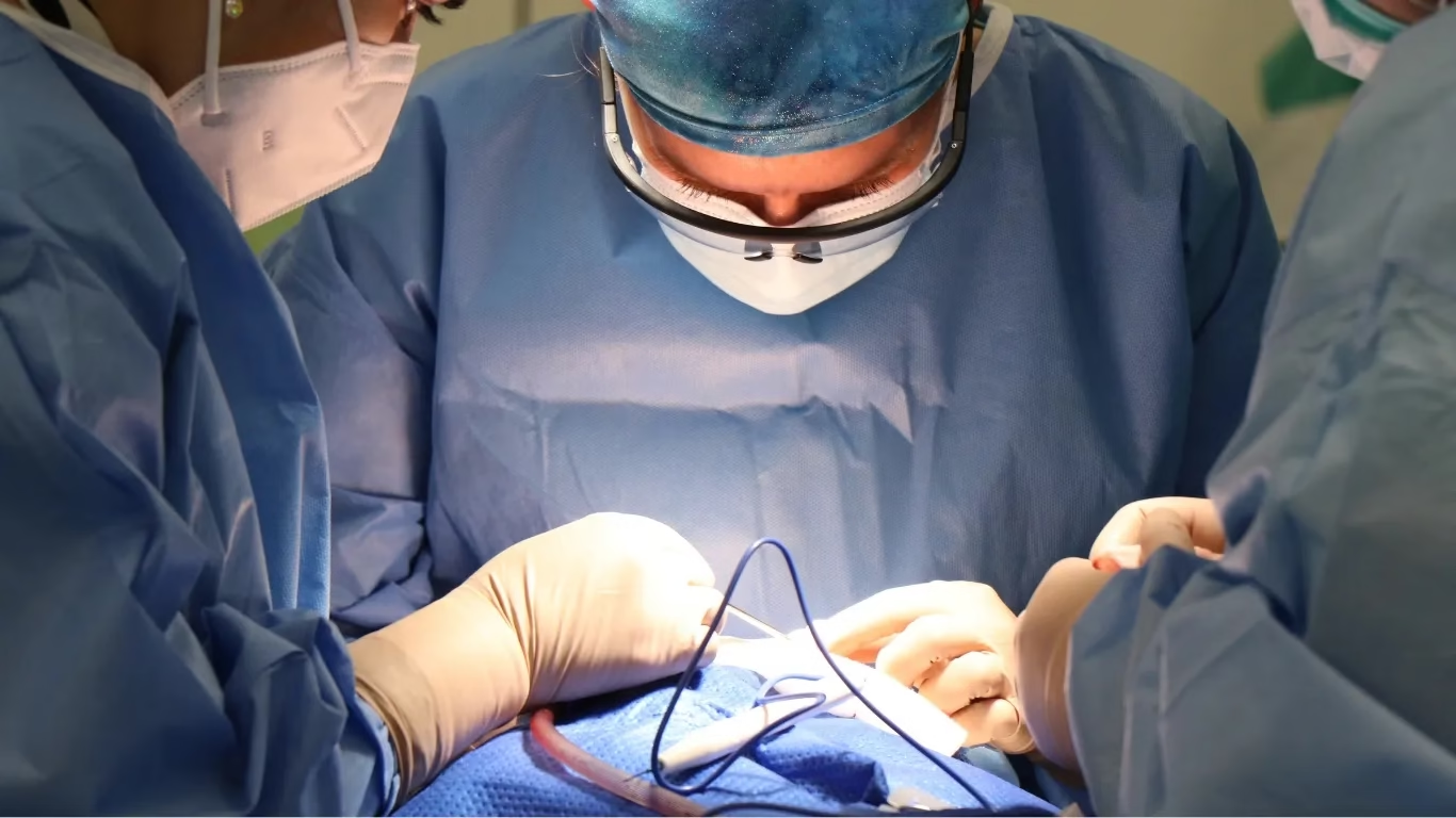
The revised technique focuses on clear identification of anatomical structures, the use of magnification when necessary, individual ligation of blood vessels and limiting excessive cauterisation near the pelvic ureter.
According to the researchers, careful dissection within this region could help minimise blood loss and reduce injuries involving the urinary tract and pelvic nerves. Preserving these structures is also important for maintaining postoperative function and patients’ quality of life.
Second Anatomical Zone Focuses on Liver Surgery
Dr Ray has also described another high-risk surgical region called “Ray’s Hepatocaval Triangle”, located near the liver and major vascular structures, including the inferior vena cava and hepatic veins.
This region is particularly challenging during liver mobilisation and dissection because of the proximity of major blood vessels. Surgical injury in this area can lead to significant bleeding and serious complications.
The proposed anatomical triangle is intended to provide surgeons with a defined reference area to guide dissection and improve awareness of nearby vascular structures.
By identifying this region before surgical manipulation, the approach aims to encourage controlled dissection and reduce the likelihood of accidental vascular injury.
Anatomical Mapping May Support Safer Oncological Procedures
Both surgical concepts emphasise the importance of recognising potentially hazardous anatomical regions before beginning dissection.
The researchers suggest that identifying these areas could support greater surgical precision, improve the preservation of surrounding organs and blood vessels, and potentially reduce complications during major cancer operations.
While the concepts offer practical anatomical guidance, further clinical evaluation would be needed to establish the extent to which they improve surgical outcomes.
The identification of these two regions highlights the role of detailed anatomical knowledge and carefully planned surgical techniques in managing complex oncological procedures.